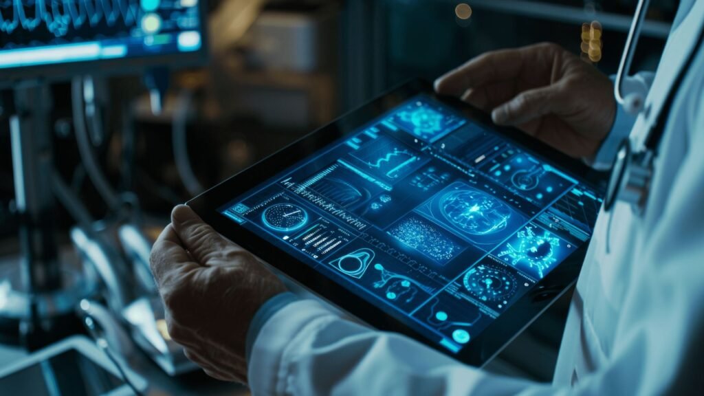
Medical imaging is a cornerstone of modern healthcare, enabling physicians to diagnose and treat conditions with a high degree of accuracy. Traditional imaging techniques like X-rays, CT scans, and MRIs provide essential insights into a patient’s condition, but computer graphics have revolutionized these techniques by improving the quality, precision, and utility of the images. By integrating advanced graphics technology, healthcare professionals can gain a better understanding of complex conditions, optimize treatment plans, and ultimately improve patient outcomes. In this article, we will explore how computer graphics enhance medical imaging, focusing on the key improvements these technologies bring to diagnosis, treatment, and medical education.

Enhanced Image Resolution and Clarity
One of the primary ways computer graphics improve medical imaging is by enhancing image resolution and clarity. High-quality images are critical in identifying conditions, determining the severity of injuries, and planning surgeries. Computer algorithms can now process medical images to sharpen details, reduce noise, and fill in missing data, making the final image more accurate and easier to interpret.
Benefits:
- Higher Image Resolution: Computer graphics can improve the resolution of low-quality images, providing a clearer view of tissues, organs, and abnormalities.
- Noise Reduction: Advanced algorithms help remove noise and artifacts from scans, leading to cleaner and more precise images.
- Better Detection: With enhanced clarity, doctors can identify small changes in tissues or organs that might otherwise go unnoticed, improving early detection of diseases such as cancer or cardiovascular conditions.
These improvements in image quality result in more accurate diagnoses and better treatment decisions.
3D Imaging and Visualization
Traditional medical images often provide a limited 2D view of a patient’s internal structures, which can be challenging when dealing with complex conditions. Computer graphics enable the conversion of 2D images into 3D models, providing a more comprehensive and accurate view of the anatomy. This 3D visualization allows healthcare professionals to gain a better understanding of spatial relationships between organs, tissues, and other structures.
Benefits:
- Better Anatomical Understanding: 3D models provide a detailed, realistic view of the human body, allowing for more accurate assessments of abnormalities and disease progression.
- Surgical Planning: Surgeons can use 3D images to visualize the exact location and orientation of tumors or other anomalies, allowing for more precise surgery planning and reduced risks.
- Patient Communication: 3D visualization also helps in explaining complex conditions to patients, improving their understanding and confidence in the treatment process.
By providing more detailed and interactive images, 3D medical imaging significantly enhances both diagnosis and treatment planning.
Real-Time Imaging and Monitoring
With the integration of computer graphics into medical imaging systems, real-time monitoring and imaging during procedures have become more effective. For instance, technologies like computer-assisted tomography (CT) and MRI can now generate real-time 3D images during surgical procedures, allowing healthcare professionals to monitor a patient’s condition live as the procedure unfolds.
Benefits:
- Guided Procedures: Surgeons and radiologists can use real-time images to guide their actions during minimally invasive procedures, such as biopsies or catheter insertions, increasing the accuracy and safety of the intervention.
- Live Updates: Real-time imaging provides immediate updates on how the body is reacting during treatments, such as monitoring the effectiveness of radiation therapy or the progress of a surgical procedure.
- Improved Accuracy: With live, continuously updated images, healthcare providers can make quick, informed decisions that enhance patient outcomes.
The ability to monitor a patient’s condition in real-time improves the overall effectiveness and safety of medical procedures.
Quantitative Imaging and Diagnostics
Computer graphics and advanced computational algorithms have enabled the creation of more sophisticated diagnostic tools in medical imaging. Quantitative imaging uses mathematical models and algorithms to extract precise measurements from images, such as tissue volume, blood flow, or the size of a tumor. This data is invaluable for making accurate diagnoses and tracking disease progression over time.
Benefits:
- Accurate Measurements: Quantitative imaging tools provide precise measurements that help doctors understand the size, shape, and density of abnormalities, leading to more accurate diagnoses.
- Objective Assessments: Unlike traditional visual assessments, quantitative imaging removes the subjectivity involved in interpreting medical images, ensuring that diagnosis and treatment decisions are based on objective data.
- Tracking Disease Progression: By comparing quantitative measurements over time, healthcare professionals can track the progress of diseases, such as cancer or degenerative conditions, and adjust treatment plans accordingly.
Quantitative imaging allows for more accurate monitoring of patients, enabling timely interventions and personalized treatment plans.
Conclusion
The integration of computer graphics into medical imaging has transformed how healthcare professionals approach diagnosis, treatment, and patient care. By enhancing image quality, enabling 3D visualization, providing real-time feedback, and improving treatment planning, computer graphics allow for more accurate and efficient medical procedures. Additionally, they support better education, faster diagnoses, and the integration of emerging technologies like AI to ensure the best possible outcomes for patients. As technology continues to advance, computer graphics will play an increasingly critical role in the future of medical imaging.


